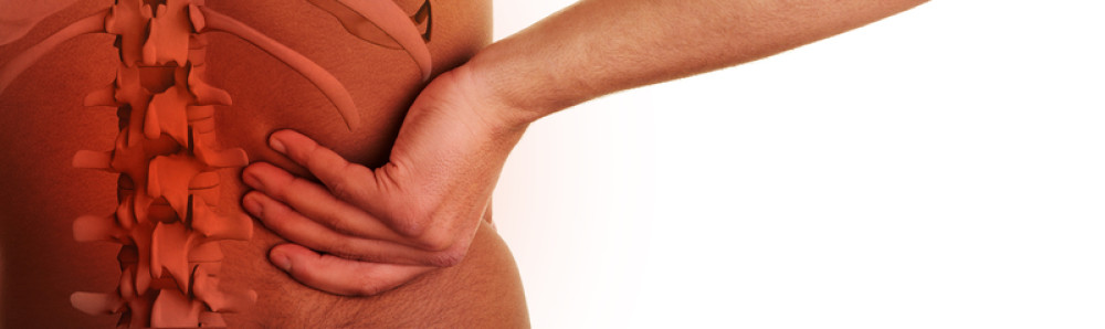
Lumbar discography involves injecting dye into the lumbar spine as an evaluation tool for those who have not been responsive to non-surgical care for low back pain. Lumbar discography helps a doctor decide whether or not to do disc surgery on the low back.
Why do a Discogram?
People have a lumbar discogram whenever they are anticipating lumbar fusion surgery. It can tell whether or not surgery will help the patient with low back pain. It is somewhat of a controversial procedure as some doctors feel the discogram is not so helpful. In truth, the helpfulness of the test depends on the skillfulness of the discographer. A bad discogram does no one any good.
This procedure can be indicated for those with groin pain, low back pain, leg pain or hip pain when other procedures haven’t been able to determine the cause of the pain. The discogram can make a difference in the diagnostic process for low back pain.
A discogram is a special test of the intervertebral discs between the vertebrae of the lumbar spine. It isn’t so much a test of what a disc looks like but instead what the disc is like physiologically. A disc that is abnormal in appearance may not hurt and may not really be abnormal physiologically. The discogram is the only test that can show what’s going on inside the disc.
While the CT myelogram and the lumbar MRI scan are great tests for the minute details of the lumbar spine but they aren’t very good at being able to tell which disc or bony part of the lumbar spine is the culprit when it comes to actual lumbar pain. It does no good to do a spinal fusion because of a “bad disc” unless pressurization of the disc reproduces the patient’s pain. As a spinal surgeon, you’d want to be able to know which disc or discs are damaged before doing an invasive procedure.
The Procedure
Every procedure begins with a history and physical to make sure that the individual is healthy enough for the procedure. The doctor who does the discogram will discuss the procedure along with its pros and cons. You’ll get a chance to ask questions of the discographer.
An IV is started and medications for sedation are given if necessary. Too much sedation makes it difficult for the patient to tell when they feel pain. The patient goes on a fluoroscopy table and ink is used to define where the injection is to be given. The back is sterilized and sterile drapes are put in a square position around the injected area. The fluoroscope is draped in a sterile fashion. Even the discographer is wearing a sterile gown.
The discographer anesthetizes a core of skin and tissue that goes to the level of the disc surface. Then a guide needle is inserted in the disc area but stops at the level of the disc. The guide needle is hollow and allows a smaller needle to enter the disc. The disc is then pressurized.
Pressurizing the Disc
This is the most important part of the discogram. Discs are pressurized using a small amount of sterile saline into the disc. During this procedure, you will be asked what you feel. You could feel absolutely nothing; you may feel pressure; you may feel pain. If you feel pain on pressurization, it can be exactly the pain you’ve been having or an unfamiliar pain that doesn’t feel like your regular pain. After this is determined, pictures by fluoroscopy might be taken, followed by a CT scan to look at the inner structure of the affected disc. The procedure can take up to an hour to do and your back will be sore for a few days, responding to Tylenol or nonsteroidal anti-inflammatory medication.
There are some possible complications of having a discogram. These include, nerve root injury, infection of the disc space and a spinal headache. The worst of these complications is the risk of disc infection because this is very difficult to treat.